Knee
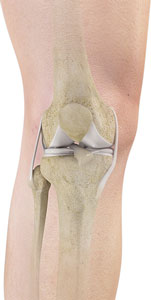
The knee is a complex joint made up of different structures including bones, tendons, ligaments and muscles. They all work together to maintain normal function and provide stability to the knee during movement.
Having a well-functioning healthy knee is essential for our mobility and ability to participate in various activities. Understanding the anatomy of the knee enhances your ability to discuss and choose the right treatment procedure for knee problems with your doctor.
Bones
The Knee is a hinge joint made up of two bones, the thigh bone (femur) and the shinbone (tibia). There are two round knobs at the end of the femur called femoral condyles which articulate with the flat surface of the tibia called the tibial plateau. The tibia plateau on the inside of the leg is called the medial tibial plateau, and on the outside of the leg it is called the lateral tibial plateau.
The two femoral condyles form a groove on the front (anterior) side of the knee called the patellofemoral groove. A small bone called the patella sits in this groove and forms the knee cap. It acts as a shield and protects the knee joint from direct trauma.
A fourth bone called the fibula is the other bone of the lower leg. This form a small joint with the tibia. This joint has very little movement and is not considered a part of the main joint of the knee.
Articular Cartilage and Menisci
Movement of the bones causes friction between the articulating surfaces. To reduce this friction, all articulating surfaces involved in movement are covered with a white, shiny, slippery layer called articular cartilage. The articulating surface of the femoral condyles, tibial plateaus and the back of the patella are covered with this cartilage. The cartilage provides a smooth surface that facilitates easy movement.
To further reduce friction between the articulating surfaces of the bones, the knee joint is lined by a synovial membrane which produces a thick clear fluid called synovial fluid. This fluid lubricates and nourishes the cartilage and bones inside the joint capsule.
Within the knee joint between the femur and tibia there are two C shaped cartilaginous structures called menisci. Menisci function to provide stability to the knee by spreading the weight of the upper body across the whole surface of the tibial plateau. The menisci help in load bearing by preventing the weight from concentrating onto a small area, which could damage the articular cartilage. The menisci also act as a cushion between the femur and tibia by absorbing the shock produced by activities such as walking, running and jumping.
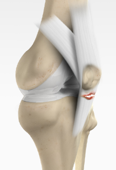
Ligaments
Ligaments are tough bands of tissue that connect one bone to another bone. The ligaments of the knee function to stabilize the knee joint. There are two important groups of ligaments that hold the bones of the knee joint together, collateral ligaments and the cruciate ligament.
Collateral ligaments are present on either side of the knee. They function to prevent the knee from moving too far during side to side motion. The collateral ligament on the inside is called the medial collateral ligament (MCL) and the collateral ligament on the outside is called the lateral collateral ligament (LCL).
Cruciate ligaments- This group of ligaments, present inside the knee joint, control the back and forth motion of the knee. The Cruciate ligament in the front of the knee is called anterior cruciate ligament or ACL and the cruciate ligament in the back of the knee is called posterior cruciate ligament or PCL.
Muscles
Muscles: There are two major muscles, the quadriceps and the hamstrings, which enable movement of the knee joint. The quadriceps muscles are in the front of the thigh. When the quadriceps muscles contract, the knee straightens. The hamstrings are in the back of the thigh. When the hamstring muscles contract, the knee bends.
Tendons
Tendons are structures that attach muscles to the bone. The quadriceps muscles of the knee meet just above the patella and attach to it through a tendon called the quadriceps tendon. The patella further attaches to the tibia through a tendon called the patella tendon. The quadriceps muscle, quadriceps tendon and patellar tendon all work together to straighten the knee. Similarly, the hamstring muscles at the back of the leg are attached to the knee joint with the hamstring tendon.
-
Knee Pain
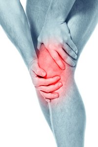
The knee is one of the largest joints in the body, formed by the lower end of the femur, upper end of the tibia and the patella or knee cap.
Read more -
Anterior Knee Pain
Anterior knee pain is a characterized by a chronic pain over the front and center of the knee joint.
Read more -
Runner’s Knee
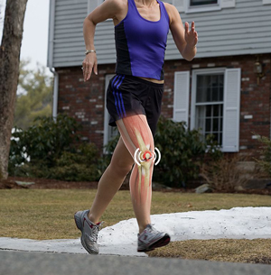
Runner's knee, also called patellofemoral pain syndrome refers to pain under and around your kneecap. Runner’s knee includes several medical conditions such as anterior knee pain syndrome,...
Read more -
Chondromalacia Patella
The patella, also called the kneecap is a small bone present on the front of your knee joint.The underside of the patella is covered by cartilage that allows smooth...
Read more -
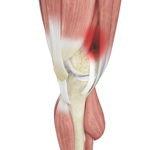
Jumper’s Knee
Jumper’s knee, also known as “patellar tendinitis" is an inflammation of the patellar tendon that connects your kneecap (patella) to your shinbone. This tendon helps in extension of the lower leg.
Read more -
Bursitis
A bursa is a small fluid-filled sac found between soft tissues and bones. It lubricates and acts as a cushion to decrease friction between bones when they move. Bursitis refers to the inflammation and swelling of the bursa.
Read more -
Baker’s Cyst
The knee consists of a fluid called synovial fluid, which reduces friction between the bones of the knee joint while you move your leg. Sometimes this fluid is produced in excess, resulting in its accumulation in the back of your knee.
Read more -
Iliotibial Band Syndrome
Iliotibial band syndrome is an overuse injury resulting from the inflammation of iliotibial band. Iliotibial band is a tough group of fibers that begins at the iliac crest of hip...
Read more -
Osteochondritis Dissecans
Osteochondritis dissecans is a joint condition in which a piece of cartilage, along with a thin layer of the bone separates from the end of the bone because of inadequate blood supply.
Read more -
Shin Splints
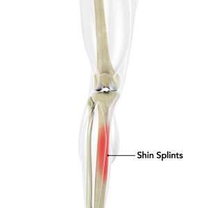
“Shin splints” is used to describe the pain and inflammation of the tendons, muscles and bone tissue around the tibia or shine bone (a large bone in the lower leg).
Launch Movie Read more -
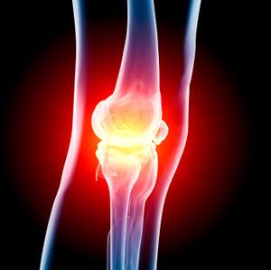
Knee Injury
Pain, swelling and stiffness are the common symptoms of any damage or injury to the knee. If care is not taken during the initial phases of injury, it may lead to joint damage that may end up destroying your knee.
Read more -
Unstable Knee
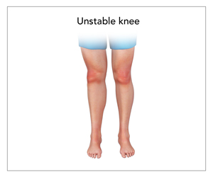
The knee joint is one of the largest joints in the body. This highly complex joint has several tissues supporting and stabilizing its movement.
Read more -
Knee Sprain
Knee sprain is a common injury that occurs from overstretching of the ligaments that support the knee joint.
Read more -
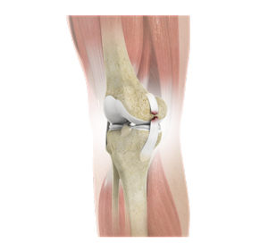
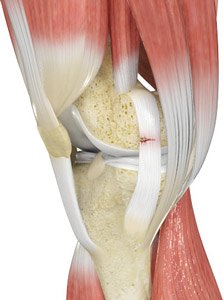
ACL Tears
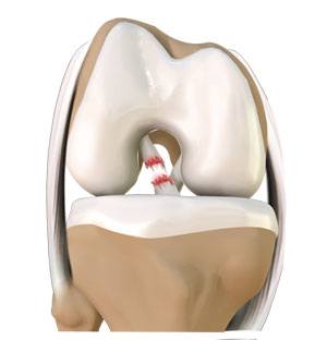
The anterior cruciate ligament, or ACL, is one of the major ligaments of the knee that is in the middle of the knee...
Read more -
MCL Tears
The medial collateral ligament (MCL) is the ligament that is located on the inner part of the knee joint. It runs from the femur (thighbone) to the top of the tibia (shinbone) and helps in stabilizing the knee.
Read more -
MCL Sprain
The medial collateral ligament (MCL), a band of tissue present on the inside of your knee joint, connects your thigh bone and shin bone (bone of your lower leg).
Read more -
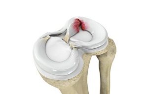
Meniscal Injuries
The knee is one of the most complex and largest joint in the body and is more susceptible to injury. Meniscal tears are one among the common injuries to the knee joint.
Read more -
Meniscal Tears
Meniscus tear is the commonest knee injury in athletes, especially those involved in contact sports.
Launch Movie Read more -
Ligament Injuries
The knee is a complex joint which consists of bone, cartilage, ligaments and tendons that make joint movements easy...
Read more -
Multiligament Instability
The knee is a complex joint of the body which is vital for movement. The four major ligaments of the knee are anterior cruciate ligament, posterior cruciate ligament, medial collateral ligament and lateral collateral ligament.
Read more -
Knee Arthritis
Arthritis is a general term covering numerous conditions where the joint surface or cartilage wears out. The joint surface is covered by a smooth articular surface...
Read more -
Patellar Dislocation/Patellofemoral Dislocation
Patella (knee cap) is a protective bone attached to the quadriceps muscles of the thigh by quadriceps tendon.
Read more -
PCL Injuries
Posterior cruciate ligament (PCL), one of four major ligaments of the knee are situated at the back of the knee. It connects the thighbone (femur) to the shinbone (tibia).
Read more -
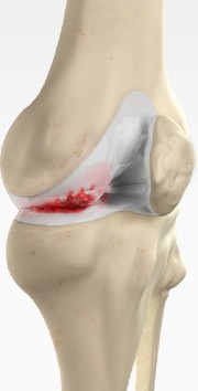
Chondral (Articular Cartilage Defects)
Articular or hyaline cartilage is the tissue lining the surface of the two bones in the knee joint. Cartilage helps the bones move smoothly against each other...
Read more -
Patellofemoral Instability
The knee can be divided into three compartments: patellofemoral, medial and lateral compartment.
Launch Movie Read more -
Quadriceps Tendon Rupture
Quadriceps tendon is a thick tissue located at the top of the kneecap. The quadriceps tendon works together with the quadriceps muscles to allow us to straighten our leg.
Read more -
Patella Tendon Rupture
Patella tendon rupture is the rupture of the tendon that connects the patella (knee cap) to the top portion of the tibia (shin bone). The patellar tendon works together...
Read more -
Lateral Meniscus Syndrome
The knee joint is formed by the union of two bones, namely the femur (thigh bone) and the tibia (lower leg bone). At the junction of these two bones is a cartilage called the meniscus, which acts as a shock absorber.
Read more -
Medial Meniscus Syndrome
Of the menisci within the knee, it is the medial that is more easily injured. Differences in the anatomical attachments of the medial meniscus compared to the lateral...
Read more
-
Platelet Rich Plasma Therapy
Our blood consists of a liquid component known as plasma. It also consists of three main solid components which include the red blood cells (RBCs), white blood cells...
Read more -
Viscosupplementation
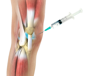
Viscosupplementation refers to the injection of a hyaluronan preparation into the joint. Hyaluronan is a natural substance present in the joint fluid that assists...
Read more -
Cortisone Injection
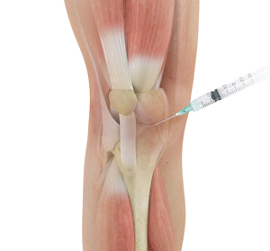
Cortisone is a corticosteroid released by the adrenal gland in response to stress and is a potent anti-inflammatory agent. Artificial preparations...
Read more -
Physiotherapy
Physiotherapy or physical therapy is an exercise program that helps you to improve movement, relieve pain, encourage blood flow for faster healing, and restore...
Read more -
Knee Arthroscopy
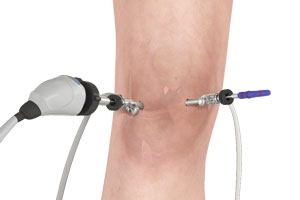
Knee Arthroscopy is a common surgical procedure performed using an arthroscope, a viewing instrument, to consider the knee joint to diagnose...
Launch Movie Read more -
Medial Patellofemoral Ligament Reconstruction
Medial patellofemoral ligament reconstruction is a surgical procedure indicated in patients with more severe patellar instability. Medial patellofemoral...
Read more -
Arthroscopic Reconstruction of the Knee for Ligament Injuries
The knee is the most complex joint in the body and is formed by the articulation between the thigh bone (femur) and the shinbone (tibia). A knee cap is present over the front...
Read more -
PCL Reconstruction
Posterior cruciate ligament (PCL), one of four major ligaments of the knee are situated at the back of the knee. It connects the thighbone (femur) to the shinbone...
Launch Movie Read more -
LCL Reconstruction
Lateral collateral ligament (LCL) is a thin set of tissues present on the outer side of the knee, connecting the thighbone (femur) to the fibula (side bone of lower leg).
Read more -
ACL Reconstruction
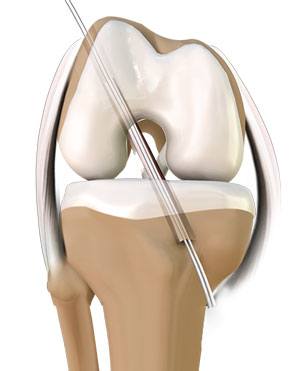
The anterior cruciate ligament is one of the major stabilizing ligaments in the knee. It is a strong rope like structure located in the center of the knee running from the femur...
Launch Movie Read more -
Multiligamentous Knee Reconstruction
Ligaments are fibrous tissue bands that connect bones and stabilize joints. The knee joint has four major ligaments – the anterior cruciate ligament, posterior...
Read more -
Patellar Tendon Repair
Patella tendon rupture is the rupture of the tendon that connects the patella (knee cap) to the top portion of the tibia (shin bone). The patellar tendon works...
Read more -
Knee Ligament Reconstruction
The knee is the most complex joint in the body and is formed by the articulation between the thigh bone (femur) and the shinbone (tibia). A knee cap is present over the front...
Read more -
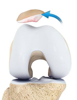
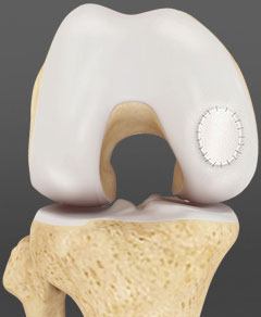
Cartilage Replacement
Cartilage replacement is a surgical procedure performed to replace the worn-out cartilage with the new cartilage. It is usually performed to treat patients...
Read more -
Cartilage Repair and Transplantation
Articular Cartilage is the white tissue lining the end of bones where these bones connect to form joints. Cartilage acts as cushioning material and helps...
Launch Movie Read more -
OATS
OATS is “osteochondral autograft transfer system”. It is one of the two types of cartilage transfer procedures and the other procedure is “Mosaicplasty”.
Launch Movie Read more
Non-surgical Treatments
Surgical Treatment
Knee Reconstruction
Cartilage Restoration
-
Autologous Chondrocyte Implantation
Autologous chondrocyte implantation (ACI) is a procedure to treat the articular cartilage defects of the knee. This procedure is effective for treating small...
Read more -
Subchondroplasty
Knee osteoarthritis (OA) is a common form of arthritis that causes joint pain and stiffness. It is a progressive disease in which the joint cartilage gradually...
Read more -
Partial Meniscectomy
Partial meniscectomy is a surgical procedure to remove the torn portion of the meniscus from the knee joint. Meniscus is the C-shaped cartilage located...
Read more -
Meniscal Surgery
Meniscus tear is the commonest knee injury in athletes, especially those involved in contact sports. A suddenly bend or twist in your knee cause the meniscus...
Launch Movie Read more -
Knee Preservation Surgery
Coming Soon...
Read more -
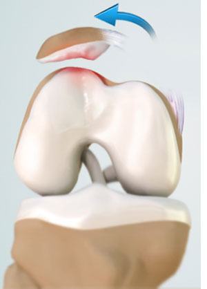
Microfracture
Microfracture is a surgical technique used to repair articular cartilage damage in the knee called chondral defects. Articular cartilage is a complex...
Read more